Find us on Facebook and Twitter

Entries by Henderson Equine Clinic (42)

Monday
Nov032014

On Farm Physical

What Every Horse Owner Should Know….


As you head out to feed your horse in the evening you notice that he/she has some dirt or shavings on their back and this morning’s hay is still on the ground.  He doesn’t seem interested in dinner and instead starts pawing and rolling on the ground. 


Its time to call your veterinarian and they may ask you to check your horse’s heart rate, gut sounds and mucous membrane color.


Knowing how to perform a basic physical examination is a skill every horse owner should know. It is also important to know your horses' normal vital signs. Horse’s have a wide range of normal values for physical exam parameters, and it is useful to compare values in an emergency to your horses' known normal values. As a general rule, the equine heart rate ranges from 32-44 beats per min (bpm), respiratory (breath) rate between 8-16 breaths per min (bpm), and normal temperature between 99.5°-101.5°F.


To take your horse's heart rate, place a stethoscope just behind their elbow on the left side of their chest. You will hear a "lub-dub", which constitutes one heart beat. You can also use their pulse to estimate their heart rate. Feel under the round aspect of the back of the jaw and you will feel a round artery and vein. Place light pressure with your fingertips until you can feel a pulse.


To take your horse's respiratory rate, you can watch the rise and fall of their chest or abdominal wall with their breaths. In normal horses it may be a subtle movement. You can also place your hand just in front of the horse’s nostril and feel for the breath on your palm, though some horses may increase their respiratory rate due to something in front of their face.


To take your horse's temperature, stand behind them and to the side. Gently lift their tail, and insert a lubed thermometer into their rectum. If you have a non digital thermometer, be sure to hold it in place or tie a string from the thermometer to their tail and leave in place for a full 2 minutes. Make sure you stand in a safe position, as some horses will kick during this procedure.


Some other parameters that are good to look at include mucous membranes. To look at a horse’s mucous membranes, gently flip their upper lip and look at their gums. Normally their gums should be light pink and moist. You also can check a capillary refill time (CRT), which is an indicator of systemic perfusion. To do this, press your thumb into their gum until the tissue underneath it blanches white. Then release the pressure and count how many seconds it takes for the pink color to return to the area. Normal CRT is less than 2 seconds.

Gastrointestinal sounds (Gut sounds) are another parameter that your veterinarian may ask you to listen to. They can vary between horses and depending on time of day and what they are being fed.  The best way to learn what your horse’s normal gut sounds are is to listen to them at least once a season (green grass gurgles, normal pasture noises, grain gas bubbles, etc). Gut sounds are particularly useful in colic cases, where gut sounds may be increased or decreased.


Digital pulses are a key parameter, particularly in laminitis or lameness cases. To feel for digital pulses, feel on the outside of the pastern about mid between the coronary band and fetlock for a group of round structures.  Palpate gently over this area to see whether you feel a pulse. It is common to not feel digital pulses in normal horses, but very strong pulses to their hooves can indicate a problem.


By performing a brief physical examination on your horse, you can provide important information to your veterinarian before they are able to reach the farm, and give some direction as to whether there are any treatments that can be initiated immediately. However, always be sure to contact a veterinarian before administering any medications or starting treatment.

Thursday
Aug072014

Navicular Syndrome

(From the researchers at Kentucky Equine Research)

Navicular syndrome (or navicular disease, or caudal heel pain syndrome) is a degenerative condition of structures in the horse's heel. The navicular bone lies at the back of the heel, and the deep digital flexor tendon runs down the leg and wraps under the navicular bone before anchoring to the coffin bone. Pain results from changes in the bones, bursa (fluid-filled joint structures designed to absorb shock and reduce friction), tendons, and ligaments in this area.


What signs does an affected horse show?

Lameness is the classic sign of navicular syndrome. This can appear suddenly, but a more common pattern is mild lameness that becomes progressively worse over time. A horse with navicular syndrome feels pain in the heels of the front feet, and its movements reflect attempts to keep pressure off this area. At rest, the more painful foot is often "pointed," or held slightly in front of the other forefoot, thus bearing little or no weight. Because the horse tries to impact the ground flat-footed or toe-first instead of the more normal heel-first pattern, the gaits are short-strided and stiff. A horse with navicular syndrome has difficulty turning sharply, going downhill, and moving on rocky or hard ground. Picking up one front foot for trimming or shoeing is painful because weight is concentrated on the other foreleg, and affected animals may become quite uncooperative during farrier visits.

Does navicular syndrome affect all types of horses?

While there's no guarantee that a particular horse will or will not develop navicular trouble, the problem is most common in stock-type horses (Quarter Horses, Paints, Appaloosas). There is a fairly high incidence in Thoroughbreds and some warmblood breeds. Arabians, on the other hand, are rarely affected. Lameness from navicular syndrome is most often diagnosed in horses between the ages of seven and fourteen.

What causes this condition?

No one knows precisely what causes navicular syndrome. Like some other lamenesses, a combination of factors is probably to blame. Conformation seems to be important, with more cases occurring in horses with heavy bodies, upright pasterns, and small hooves. A large number of affected horses have a history of work involving front-leg impact (jumping, cutting, roping, and reining) or increased concussion (parade work or other use involving hard or rocky surfaces). Suspicion has also been directed at irregular farrier care, unbalanced hooves, and shoeing practices that reduce contact between the frog and the ground. A common thread seems to be the combination of increased stress and limited oxygenation of structures in the heel area, but the exact cause of tissue damage and inflammation has not yet been determined.

How is the diagnosis made?

Heel pain is not always caused by navicular disease: temporary lameness from bone fractures, muscle strains, and trauma to tendons or ligaments can mimic the syndrome. A vet may need to use a combination of flexion tests, hoof and frog pressure tests, nerve blocks, X-rays, scintigraphy, thermography, ultrasound, venograms, and magnetic resonance imaging (MRI) to determine the cause of the horse's discomfort. Diagnosis is made after a consideration of the horse's history, use, conformation, and test results.


At one time the appearance of lollipop-shaped invaginations (holes) in the navicular bone on X-rays was considered to be definitive evidence of navicular syndrome. However, it has been found that not all horses with such invaginations exhibit clinical signs of lameness, and some acutely lame horses fail to show any trace of bone abnormality. Interestingly, research at the Idaho Museum of Natural History found lollipops in 17% of navicular bones recovered from equine skeletons up to three million years old, showing that the phenomenon is not of strictly modern origin. In the "history repeats itself" category, bones from large-bodied horses were more likely to show invaginations, and animals living in areas of hard and rocky terrain were also more often affected than grassland-dwelling animals.

Can navicular syndrome be cured?

Consultations with a veterinarian and a farrier are the first steps in combating navicular syndrome. While there is no cure, a prompt diagnosis allows treatment-farrier, medical, or surgical-to begin early in the course of the disease. Proper trimming and therapeutic shoeing can provide pain relief for many horses. Farrier care is aimed at correcting broken-back or broken-forward pastern angles and normalizing underrun or contracted heels. Generally a shortened toe, either through trimming or shoe design, is a goal. Heel support afforded by egg bar or wide-web shoes reduces pain in some horses, and a shock-absorbing polyurethane shoe from Switzerland is being tested as a possible therapeutic aid. Overall, proper trimming and shoeing can relieve discomfort in about 30% of horses with navicular syndrome.

Polysulfated glycosaminoglycans and hyaluronic acid sometimes lead to improvement, possibly by inhibiting enzymes involved in tissue breakdown. Anti-inflammatory medications can be injected into the heel area or given orally for pain relief. Medical treatment combined with therapeutic shoeing can help about 60% of affected horses.

Palmar digital neurectomy, a surgical procedure to sever the nerves to the painful area, is a last resort in treating navicular syndrome. This option eliminates sensation in the rear third of the foot, thus ending pain and lameness, but it is not a permanent cure. Degenerative changes continue to occur within the hoof, and about one-third of treated horses are lame again within two years.

Can feeding practices help to prevent or treat navicular syndrome?

Navicular syndrome is not directly caused by feeding practices, but as with any condition affecting the legs and feet, an overweight horse puts excessive strain on its musculoskeletal system. With the strong correlation between heavy-bodied, small-footed horses and navicular syndrome, common sense should warn owners not to allow their horses to become too fat. 

Pasture intake can be limited by muzzling or dry-lotting horses, and easy-keeping animals can be fed a low-calorie supplement pellet such as All-Phase, thus providing essential vitamins and minerals without an overabundance of calories. Increasing exercise is also beneficial for keeping horses in trim condition. Although turnout and light exercise are preferred to stall rest for navicular horses, heavy work is usually ruled out as a conditioning measure for these animals. Keeping a horse at a mid-range body weight may delay the onset of navicular syndrome in susceptible individuals, and can also help to keep affected animals more comfortable.


Friday
Jul112014

Equine Protozoal Myeloencephalitis (EPM)

Equine Protozoal Myeloencephalitis (EPM) is a master of disguise. This is a serious disease, which attacks the horse’s central nervous system.  It can be difficult to diagnose because its signs often mimic other health problems in the horse and signs can range from mild to severe.

EPM is caused by one of two protozoal organisms, Sarcocystis neurona or Neospora hughesi, though almost all infections in the United States are caused by S. neurona. More than 50 percent of all U.S. horses have been exposed to S. neurona, which is carried by the opossum and spread through hay, water and pasture contaminated with opossum feces.  Fortunately, not all horses exposed to the parasite develop the disease. Many horses are able to mount an immune response to the protozoa before it is able to affect the central nervous system.  These horses will carry the antibodies generated by this immune response for life.  Future diagnostic testing will read these antibodies as a positive sign of the disease, complicating the interpretation of the test results. Researchers are unable to identify why some horses are able to successfully fight the protozoa and some are not.


If the protozoa are able to cross the blood-brain barrier and enter the central nervous system, they live inside cells in the central nervous system and are not able to be attacked by the immune system.  Changes in the body’s chemistry typically caused by significant or stressful events (trailering, high levels of athletic performance, injury, pregnancy, poor nutrition/weight loss, etc) cause the protozoa to reproduce and the stressed immune system cannot stop their spread into other areas of the brain or spinal cord.  As they spread they cause lesions and inflammation that cause the clinical signs of EPM.  The amount of time from horse consuming the protozoa and clinical disease can be anywhere from weeks to two years.


The clinical signs of EPM can be quite varied, depending on the severity and location of the lesions that develop in the brain, brain stem or spinal cord. Clinical signs are usually asymmetrical (not the same on both sides of the horse). The American Association of Equine Practitioners (AAEP) uses the following checklist of symptoms from when assessing a  horse’s condition for the possibility of EPM:


  • Ataxia (incoordination), spasticity (stiff, stilted movements), abnormal gait or lameness.

  • Incoordination and weakness which worsens when going up or down hills or when head is elevated.

  • Muscle atrophy, most noticeable along the topline or in the large muscles of the hindquarters, but can sometimes involve the muscles of the face or front limbs.

  • Paralysis of muscles of the eyes, face or mouth, evident by drooping eyes, ears or lips.

  • Difficulty swallowing.

  • Abnormal sweating.

  • Loss of sensation along the face, neck or body.

  • Head tilt with poor balance; horse may assume a splay-footed stance or lean against stall walls for support.

  • Seizures or collapse.


If left undiagnosed and untreated, EPM can cause devastating and lasting neurological damage.  Contact your veterinarian immediately if you suspect your horse has developed signs of a neurological disease.  The sooner a diagnosis is made and treatment begins, the better the horse’s chances for recovery.  For more information on methods of prevention and the treatment options (there are many options, opinions, and varied results for all), contact your veterinarian.


Friday
May302014

When Mosquitoes Go Bad part 1.......(West Nile Virus)

WHEN MOSQUITOES GO BAD, PART 1

West Nile Virus

When you hear about West Nile Virus, it brings to mind images of dead crows and mosquito laden swamps.  In the past few years, West Nile Virus (WNV) has been diagnosed in multiple horses in Western New York.  Last year (2013) The New York Department of Agriculture and Markets reported 13 WNV positive horses throughout the state!


While the immune system of most horses infected by WNV can prevent the virus from crossing the blood-brain barrier, clinical signs are seen in some horses when the virus breaches the blood-brain barrier and causes encephalomyelitis, damaging the brain and spinal cord.  The clinical signs of WNV encephalomyelitis vary in range and severity, with the most frequently observed signs include incoordination or ataxia (especially of the hind limbs); twitching of the muzzle and lower lip, and twitching of the muscles in the neck, shoulders or pectoral (chest) region. Signs may be the same on the left and the right of the horse or may be one sided. Also reported are behavioral abnormalities such as depression or heightened sensitivity to external stimuli, stumbling, toe dragging, leaning to one side and in severe cases, paralysis of the hindquarters, recumbency, coma and death. Other clinical signs that may be noted include fever, generalized weakness, impaired vision, inability to swallow, aimless wandering, and convulsions. The nature and severity of clinical signs depend largely on the area(s) of the central nervous system affected by the virus and the extent of damage. There are reports of WNV occurring more commonly in older horses; however, last years cases in New York ranged from yearlings to aged horses.   

First isolated in Uganda in 1937, WNV is transmitted principally by mosquitoes and can cause inflammation of the brain and spinal cord (encephalomyelitis). Clinical disease caused by this virus is seen primarily in birds, equines and humans and very infrequently in goats, sheep, dogs, llamas, various reptiles and bears, among other species. Prior to its discovery in the northeastern U.S. in 1999, WNV was widely distributed in Africa, the Middle East, southwest Asia, and parts of Europe. 

WNV was first recognized in the western hemisphere in September 1999, when it was isolated from the tissues of sick flamingoes and pheasants at the Bronx Zoo and from dead crows in the New York City area. By 2002 over 15,000 horses were diagnosed with WNV in 41 states.

WNV circulates in nature between birds and mosquitoes. Various species of birds serve as hosts of the virus, allowing it to replicate within them.  Mosquitoes act as vectors of WNV by biting the infected birds and then transmitting it to a wide range of animals, including humans. The strains of WNV present in North America also cause disease in crows and blue jays, in which the infection is usually fatal. Humans, horses, and a wide variety of other species can also be infected with WNV.  Because there is only a very small amount of the virus in the blood of infected horses, mosquitoes are unable to transmit the virus from horse to horse or from horse to human. The virus is transmitted when a mosquito takes a blood meal from an infected bird and then feeds on a horse. During the process of taking a blood meal from the horse, the virus is transmitted by the infected mosquito.

As is typical of numerous other viral infections, many horses experience no clinical illness following exposure to the virus for the first time. 

WNV infection diagnosis is usually based on the nature of the clinical signs displayed by an affected horse, together with the detection of antibodies to the virus in the blood by laboratory examination. It is important to remember that many of the clinical signs of WNV encephalomyelitis closely resemble those of many other equine neurological diseases (e.g., Eastern equine encephalitis, rabies, equine protozoal myeloencephalitis, equine herpesvirus-1 and botulism).  Testing and progression of clinical signs will help to differentiate between these diseases. However, cases of WNV encephalomyelitis tend to occur during late summer or early fall when viremia (the level of infective virus in the blood) in the bird population is higher and mosquito populations are numerous and active.

Treatment

Currently, there is no specific anti-viral treatment for WNV encephalomyelitis. Treatment focuses on controlling pain and inflammation. Anti-inflammatory drugs will be provided to control inflammatory changes in the central nervous system. Other supportive measures such as intravenous fluids, sedatives and nutritional support can be important components of therapy. It is important to consult your veterinarian immediately if you suspect your horse is showing neurological signs so that the appropriate treatment measures can be implemented without delay.

Prevention

A number of measures can be taken to help protect your horse against WNV. These are comprised of vaccinating against the disease coupled with management strategies to reduce exposure to mosquitoes and. Horses vaccinated against Eastern, Western or Venezuelan equine encephalomyelitis are not protected against WNV.   A separate WNV vaccine is currently available as well as combination products that combine Eastern, Western, Tetanus as well as West Nile Virus.

The vaccine should be administered as a series of two doses given three to six weeks apart. Foals should receive three immunizations starting at 6 months of age if the mare was immunized against WNV 30 days prior to foaling. The duration of immunity from vaccination is not known. It is recommended to vaccinate every four to six months in regions where the virus is active. Contact your veterinarian for the appropriate vaccination schedule for your location.  In New York we typically recommend bi-annual vaccination against WNV in the spring and a booster during the early fall months.

Aside from vaccination against WNV, other measures should be taken to reduce the risk of your horse being bitten by a virus-infected mosquito. Eliminate or reduce potential mosquito breeding sites by disposing of old receptacles, such as tires and containers and eliminating areas of standing water in areas where horses congregate. 

Clean clogged roof gutters and turn over plastic wading pools or wheelbarrows when not in use. Thoroughly clean livestock watering troughs at least monthly. Screen stalls (if possible) or at least install fans over the horses to help deter mosquitoes. Avoid turning on lights inside the stable during the evening or overnight. Because mosquitoes are attracted to light, placing incandescent bulbs around the perimeter of the stable will attract mosquitoes away from the horses. Lights can also be used to draw mosquitoes to electric bug zappers. 

The use of insect repellant that contains pyrethrin on horses can also reduce the chance of being bitten by mosquitoes. Remove any birds (including chickens) located in or close to a stable. Some veterinarians have success by hanging cattle ear tags on horse halters. These ear tags have been impregnated with insecticide and often reduce the effects of not only mosquito biting, but also midges and the effects of “fly-strike” dermatitis around the ears.  Our Mosquito and Fly Control Measures blog post has additional information on vector control!

Because WNV can affect humans as well as horses, don’t forget to take actions to protect yourself as well. When outdoors in the evening, wear clothing that covers your skin and apply plenty of mosquito repellent.

Prevention is key to the control of this infection.


Monday
May192014

Recognizing the Signs of Laminitis

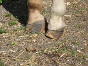
Spring is a time when veterinarians begin to see increased cases of laminitis, a painful disease that affects the feet of horses. Laminitis results from the disruption of blood flow to the sensitive and insensitive laminae within the foot, which secure the coffin bone (P3) to the hoof wall. While the exact mechanisms by which the feet are damaged is still being determined, certain precipitating events can produce laminitis. Although laminitis occurs in the feet, the underlying cause is often a disturbance elsewhere in the horse’s body.

Recent research has led to the description of the laminae in a horse’s foot as a ‘shock organ.’ That is, an organ that fails related to some sort of systemic disease. In people, the lung is considered a “shock organ”, and they are finding that the laminae in a horse’s foot reacts very similarly. This suggests that treating other diseases more effectively — like colitis, or a mare with a retained placenta, or a horse with pleurao pneumonia — we can better protect the foot from becoming the shock organ that ultimately fails and results in a crippling disease.

Laminitis can also occur after a carbohydrate overload, where your horse eats to much rich young grass or grain. An injury or trauma that causes the horse to bear less weight on one leg may also induce laminitis, as in the case of Barbaro. The continuous increased stress placed on the weight bearing limb strains the lamina that hold the coffin bone stable in the hoof, resulting in inflammation and laminitis. It is important to provide support and cushioning to all feet and legs for a horse being treated for any injury or lameness that changes how it bears weight.

Researchers have also identified a “Pre-Laminitic Metabolic Syndrome (PLMS)” in horses similar to the metabolic syndromes in people that are considered risk factors for Type II diabetes or coronary heart disease. These horses are especially sensitive to carbohydrate overload and can ‘founder’ for no apparent reason. The researchers have used cut off points for Insulin Sensitivity, Pancreatic Beta cell Response (Insulin levels compared to glucose levels), Body Condition Score and Serum Triglyceride levels to identify these PLMS ponies which are at a higher risk of developing laminitis. Identification of PLMS in ponies (and possibly horses) would allow owners and veterinarians to manage nutrition and feeding practices appropriately to prevent laminitis prior to clinical disease.

As a horse owner, it is important to recognize the signs of laminitis and seek veterinary help immediately. Signs of acute laminitis include the following:

  • Lameness, especially when a horse is turning in circles; shifting lameness when standing

  • Heat in the feet

  • Increased digital pulse in the feet

  • Pain in the toe region when pressure is applied with hoof testers

  • Reluctant or hesitant gait, as if “walking on eggshells”

  • A “rocking horse stance” in the front, with the front feet stretched out in front to alleviate pressure on the toes

Signs of chronic laminitis may include the following:

  • Rings in hoof wall that become wider as they are followed from toe to heel

  • Dished hooves, which are the result of unequal rates of hoof growth

  • Thick, “cresty” neck and abnormal fat deposits (signs of metabolic disorders that often cause laminitis)

  • Bruised soles or “stone bruises”

  • Widened white line, commonly called “seedy toe,” with occurrence of blood pockets

and/or abscesses

  • Dropped soles or flat feet


If you suspect laminitis or your horse has gotten more than a normal amount of carbohydrates (grass or grain), consider it a medical emergency and notify your veterinarian immediately. Early intervention in acute laminitis carries the best prognosis. The sooner treatment begins, the better the chance for recovery.

Your veterinarian may recommend icing your horse’s feet if it is an acute case, and placing him on deep, soft bedding with no or limited grain and lower quality hay until they are able to see them. They may also recommend applying cushion/support to your horse’s feet via boots or pads made of foam insulation sheets and duct tape.

Treating laminitis requires a multi-modal approach. Your veterinarian will need to work closely with your farrier to develop a plan and identify any changes to the coffin bone-hoof structure. X-rays may be needed to look for rotation and/or sinking of the coffin bone. Close communication between your veterinarian, farrier and yourself is essential to prevent any further changes from occurring while keeping your horse comfortable during recovery.